
Irreversible Electroporation of Metastatic Malignancy in the Liver
A 75-year-old male presented who was diagnosed in 2010 with sigmoid colon adenocarcinoma. His history included a low anterior resection of the mass in the distal sigmoid colon in January 2011 followed by 12 cycles of FOLFOX chemotherapy. In January 2012, an elevated level of carcinoembryonic antigen (CEA) and positron emission tomography/computed tomography (PET/CT) demonstrated a 3.5-cm segment VIII liver lesion. In March 2012 the patient received FOLFIRI chemotherapy with cetuximab (Erbitux).
Diagnostic Imaging
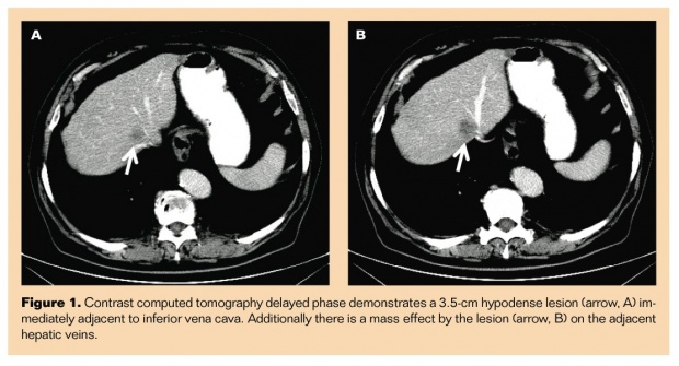
A delayed phase contrast CT demonstrated a 3.5-cm hypodense lesion (arrow, Figure 1A) immediately adjacent to the inferior vena cava.
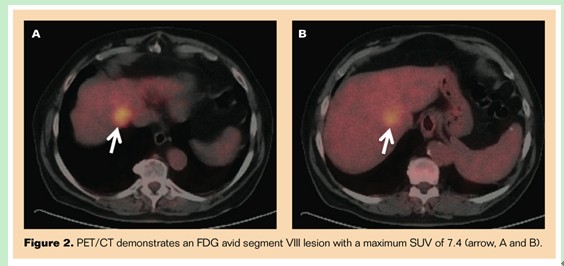
Additionally there was a mass effect by the lesion (arrow, Figure 1B) on the adjacent hepatic veins. The PET/CT demonstrated a fluorodeoxyglucose-avid segment VIII lesion with a maximum standardized uptake value of 7.4 (arrows, Figure 2).
Treatment
The patient
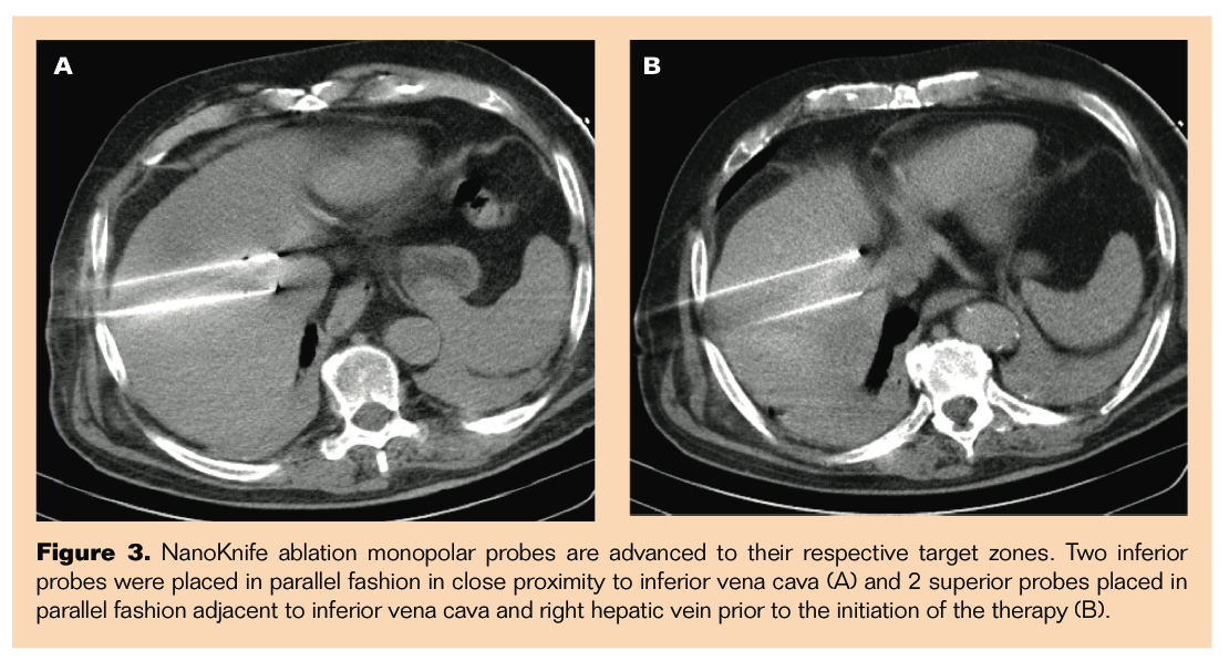
underwent irreversible electroporation (IRE) treatment with NanoKnife
(AngioDynamics). NanoKnife ablation monopolar probes were advanced to their respective target
zones. Figure 3A demonstrates 2 inferior probes placed in parallel fashion in
close proximity to the inferior vena cava (IVC), and Figure 3B demonstrates 2
superior probes placed in parallel fashion adjacent to IVC and right hepatic
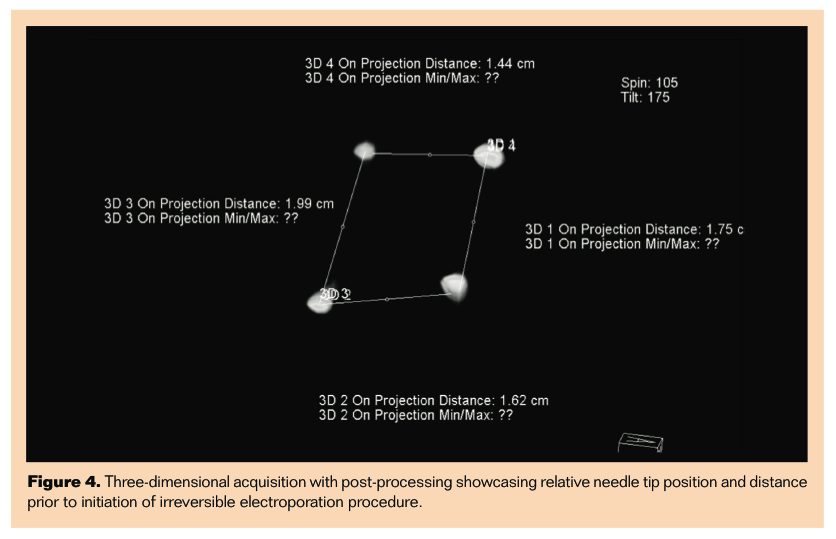
vein prior to the initiation of the therapy. Figure 4 shows 3-dimensional
acquisition with postprocessing showcasing relative needle tip position and
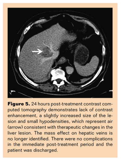
distance prior to initiation of IRE procedure. Contrast CT 24 hours post
treatment demonstrated a lack of contrast enhancement, a slightly increased
size of the lesion and small hypodensities, which represent air (arrow, Figure
5) consistent with therapeutic changes in the liver lesion. The mass effect on
the hepatic veins was no longer identified. There were no complications in the immediate post-treatment
period and the patient was discharged.
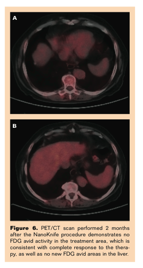
A
PET/CT scan performed 2 months after the NanoKnife procedure demonstrates no
FDG avid activity in the treatment area, which is consistent with complete
response to the therapy, as well as no new fluorodeoxyglucose-avid areas in the
liver (Figures 6A and B).
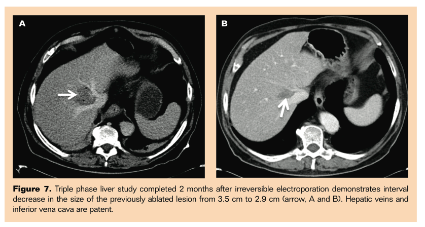
A triple phase
liver study completed 2 months after the IRE demonstrated interval decrease in
the size of the previously ablated lesion from 3.5cm to 2.9cm (arrows, Figures
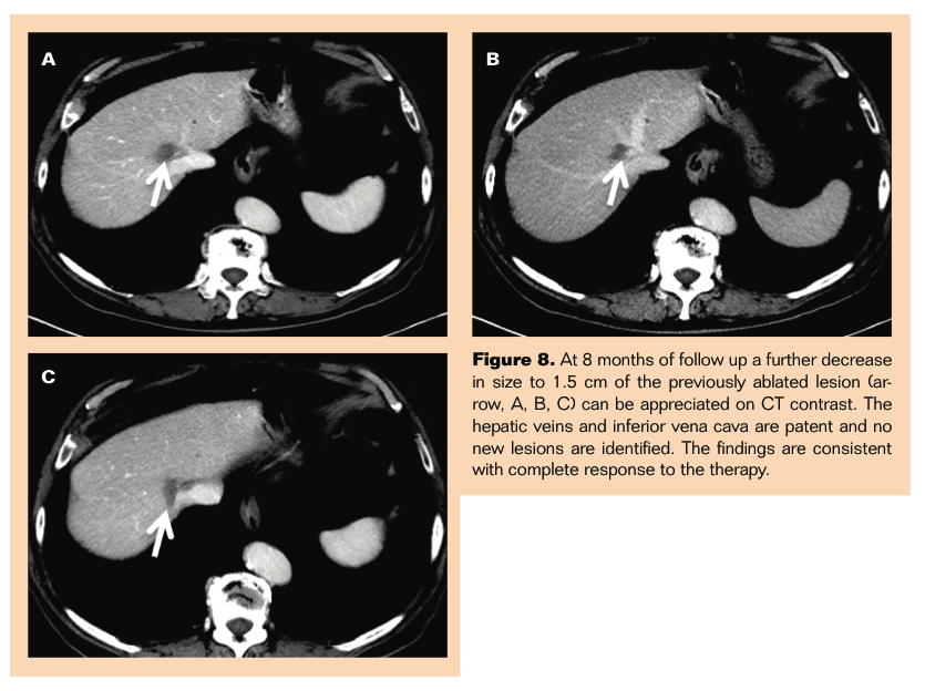
7A and B). Hepatic veins and IVC were patent. At 8 months of follow-up a
further decrease in size to 1.5 cm of the
previously ablated lesion (arrows, Figure 8) was appreciated on CT
contrast. The hepatic veins and IVC were patent and no new lesions were
identified. The findings are consistent with complete response to the therapy.
來源:Irreversible Electroporation of Metastatic Malignancy in the Liver
Issam Kably, MD1, Govindarajan Narayanan, MD2, Dmitriy Meshkov, BS3
From the 1University of Miami Miller School of Medicine--Jackson Memorial Hospital; the 2Sylvester Comprehensive Cancer Center, Miami, Florida; and 3University of Texas Medical School at Houston
 Follow on Facebook
 Follow on Twitter
 Subscribe to RSS